Eczema, Atopic Dermatitis & Topical Steroid Withdrawal (Part 1)
This article is part one in a two-part article series. Part two will discuss a case study showing progressive photos over a five month treatment period along with a discussion on the TCM herbs used for treatment. Part two will be published on April 16, 2024.
Introduction
In recent years, there has been a rise in atopic dermatitis (AD) as well as misuse or overuse of the primary treatment, which is topical corticosteroids. Complications from topical corticosteroids, including those arising from the discontinuation of these medications, can lead to significant deterioration in the the skin and lives of some individuals, and this has come to be known as “topical steroid withdrawal” (TSW).
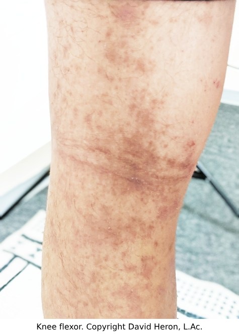
Chinese Medicine has recognized atopic dermatitis for about a millenia, naming it Si Wan Feng - Wind Of The Four Crooks, which refers to the elbow and knee flexures where itchy inflamed skin (Wind) presents most typically in patients with AD. Likewise in the West, as documented by Lio (2014), AD lesions have been documented for some time, even though a more definitive disease definition linking it to an allergic tendency is more recent.
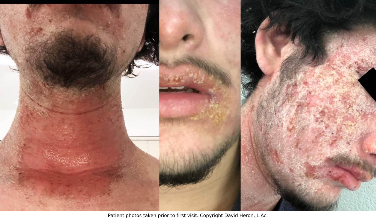
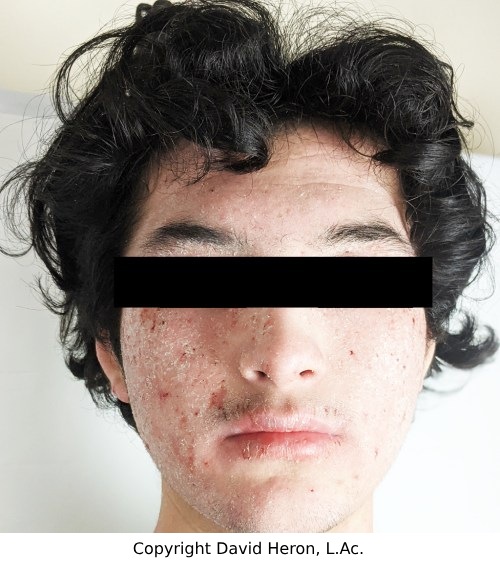
My heart has a special place for severe eczematous patients and their families. On a fairly regular basis, I meet a new patient seeking treatment for eczema / atopic dermatitis and "topical steroid withdrawal". At the first visit I notice redness on their faces, sometimes with swollen eyes, sometimes with large areas of scaling, and often with peeling skin. They are often in loose clothes to decrease pain from chronically inflamed skin. Over the course of our interview, they share their health journey and at some point they ask, with both hope and lingering doubt from previous experiences, if Chinese Medicine can significantly improve their condition? The answer is yes. I have had the good fortune to have clinically directed enough positive transformations to confidently say that both atopic dermatitis and/or topical steroid withdrawal respond well to Chinese Medicine. In part II of this article, I will describe the treatment of one such patient: an 18 year old male named M.J who sought treatment in October 2021 for "topical steroid withdrawal" that developed secondarily to the treatment of atopic dermatitis.
What is Atopic Dermatitis?
Before a discussion of TSW, it is appropriate to outline all the terms involved in order to have a clear understanding of what is meant by eczema or atopic dermatitis, and what place eczema has in a connected landscape of various skin and allergic conditions. Is eczema the same as atopic dermatitis?
Eczema is a broad term used to describe numerous related but different conditions involving redness, inflammation, itching of the skin, possibly scaling and occasionally the presence of vesicles. Each type of eczema has different causes, symptoms, and treatments. In common parlance, when people speak of eczema they are most often referring to atopic eczema, also known as atopic dermatitis. Dermatitis is another term that is used interchangeably with eczema for conditions involving skin inflammation and redness. Despite similarities in meaning, certain diseases are only used with either eczema or dermatitis in their title.

As an umbrella term, both eczema and/or dermatitis can refer to the following loosely related but different skin conditions: seborrheic dermatitis; perioral dermatitis; nummular eczema; pompholyx eczema / dyshidrotic eczema; stasis eczema; and contact dermatitis. Chinese Medicine can be utilized to treat all of these conditions. However, the rest of this article will only focus on atopic dermatitis that is further complicated with topical steroid withdrawal.
Atopic dermatitis presents as generalized dry skin, itch, and rash that often affects the knee and elbow flexures (in children and adults) but is not limited to those areas. It is also marked by a tendency to flare up, go into remission, and relapse into a flare up again (Stanway et al, 2023). The acute flare ups present with erythematous (red) and inflamed skin that is itchy. There may be some weeping and exudation of fluid. Occasionally, there can also be vesicles. After the active cycle heals, there may be some hyperpigmention or hypopigmentation along with lichenified skin, particularly after many cycles of flare up. Lichenification is the skin's response to frequent scratching and rubbing, where the skin becomes rough and often displays exaggerated skin lines.
Atopic dermatitis also refers to eczema that is secondary to "atopy" or allergy. Johansson et al. (2003) defined atopy as "a personal and/or familial tendency, usually in childhood or adolescence, to become sensitized and produce IgE antibodies in response to ordinary exposures to allergens, usually proteins. As a consequence, these persons can develop typical symptoms of asthma, rhinoconjunctivitis, or eczema." The term “atopic march” describes a tendency for some individuals to develop AD in infancy and to then develop allergic rhinitis and asthma later in childhood (Bantz et al, 2014). In an earlier study performed by Gustafsson et al (2000), 94 children aged 4 to 35 months old and diagnosed with atopic dermatitis lasting at least 3 months were regularly monitored until age 7. At the conclusion of the study, 45% were found to have developed allergic rhinitis and 43% were found to have developed asthma. While it is not clear whether atopic dermatitis is necessary to develop the other atopic conditions, there is evidence that those who suffer one atopic disease are more likely to develop another. A recent study by Gabryszewski et al (2023) produced further evidence for the atopic march by analyzing the electronic health records of roughly 219,000 children; noting a tendency toward co-morbidity amongst children with atopic dermatitis, asthma, allergic rhinitis, IgE mediated food allergies, and eosinophilic esophagitis.
Why might atopic dermatitis as an infant or child lead to other allergic diseases? One common theory is the observed impairment of the skin barrier in patients with AD. These patients are often found to have different lipid compositions in the top layer of the epidermis, the stratum corneum, which does not allow the skin to retain moisture properly or for a proper barrier to develop (Hadi et al, 2021). In addition it has been found that approx 50% of atopic dermatitis patients suffer a genetic variant that leads to filaggrin deficiency (Bantz et al, 2014). Filaggrin is protein in the stratum corneum which helps create a barrier and promote proper hydration in the skin. Therefore, it is believed that due to an impaired skin barrier, microscopic antigens or airborne allergens are able to penetrate the skin, leading to immune sensitization that will later trigger an allergic response with future exposures to those allergens.
Who Develops Atopic Dermatitis?
Atopic dermatitis is the most common inflammatory skin condition in the world. It is prevalent in greater than 15% of the world's population and has been steadily growing as countries undergo greater industrial development and prosperity. According to data from the National Eczema Association (n.d.), atopic dermatitis is a significant concern among the pediatric population, with approximately 9.6 million children in the U.S. suffering from it. Alarmingly, a third of these cases present as moderate to severe cases. Since 1997, the prevalence of atopic dermatitis in children has seen an upsurge, increasing from 8% to 15% in the U.S. However, atopic dermatitis is not confined to pediatrics, as 16.5 million U.S. adults, which equates to 7.3% of the adult population, are living with this condition, with almost 40% of these cases being moderate to severe. AD generally makes its first appearance early in life, with 80% of those affected experiencing symptoms before they reach six years old. Yet, the condition isn't exclusive to early onset, with one in four adults reporting the onset of symptoms in adulthood, with 6% reporting symptoms after age 60.
Atopic dermatitis affects those of every race. Silverberg et al (2013) examined allergy rates in foreign born Americans and found that those born outside the U.S have approximately a 50% reduced chance of developing atopic dermatitis. However, after being in the U.S for one decade, the study found that the rates of allergies, including atopic dermatitis, was significantly greater than those that lived in the U.S. for 2 years or less.
How Does Atopic Dermatitis/ Eczema Present?
While atopic dermatitis can occur at any age, we typically see the initial presentation in infancy, often with seborrheic dermatitis of the scalp, axilla, and groin area (Stanway et al, 2023). Unlike cradle cap, which involves a non-inflammatory, non-itchy yellow scaling of the scalp, atopic dermatitis involves an inflammatory rash that often spreads to the face, especially the cheeks and often the dorsum of the hands. Fortunately, Chinese Medicine offers some relatively easy topical treatments for infants that will be discussed further below.
As children progress in age, the location of the eczema lesions starts to move toward the more typical location of the elbow and knee flexures but is in no way limited to those areas. The neck is another area often affected by eczema, and this is often referred to as “dirty neck syndrome.” The lower eyelids are often affected and complicated by rubbing. As children develop a greater ability to scratch, lichenification usually develops in highly pruritic areas. If AD continues into adulthood, it can also present in varying forms with papular and discoid eruptions, significant lichenification, and loss of or thinning of the eyebrows from inflammation and rubbing and / or scratching (Stanway et al, 2023).
There are numerous possible complications, such as infections from the impaired skin barrier function and the abnormal immune response inherent to eczema (Stanway et al., 2021). Common infections can include infection with staphylococcus aureus as typically seen in impetigo, molluscum contagiosum, and eczema herpaticum. Fungal infections of the skin are also more common in those with eczema.
Another complication is hyper- and hypopigmentation. While these are harmless to the patient's physical health, lingering discoloration of the skin can add a further burden to some patient's mental health. Both hypopigmentation and hyperpigmentation can come as a result of the dermatitis itself, however improper use of steroids can also lead to more enduring and even permanent hypopigmentation.

Erythroderma is a complication that refers to a widespread and intense reddening of the skin (erythema) due to inflammation. Erythroderma often precedes exfoliation or peeling of the skin (Ngan et al., 2016), and can occur in various skin diseases, including AD. Erythroderma is a common and core aspect of topical steroid withdrawal.
What is a Topical Steroid or a Topical Corticosteroid?
Corticosteroids are a class of steroid hormones that are produced in our adrenal cortex. Endogenous corticosteroids, which are naturally produced by the body, play critical roles in a variety of physiological processes, including the regulation of inflammation, immune response, metabolism, and maintaining homeostasis under stress conditions. Due to their ability to reduce inflammation and reduce over activity of the immune system, they have been useful as medications. Topical corticosteroids are applied to the skin to help reduce inflammation and immune response and they also cause vasoconstriction of blood vessels which can help reduce erythema, where vasodilation plays a role. They have been commonly used to treat conditions like atopic dermatitis, psoriasis, and various other types of dermatitis amongst others. These medications come in various strengths, from mild to very potent, and should be used under the guidance of a healthcare provider to minimize potential side effects. In the United States, topical steroids are often grouped into 7 categories; ranked from strongest (Class I) to weakest (Class VII). For reference, see Gabros et al (2023) and Ference et al (2009).
Recommended Guidelines and Possible Side Effects of Topical Steroids
It should be noted that steroid medications, including topical corticosteroids, have been a major medical breakthrough that regularly saves lives. However, due to their potent nature, they must be used appropriately and thoughtfully.
Much of the literature states that topical corticosteroids (TCS) are generally safe when used short-term and certain guidelines are followed and that side-effects are rare. Ference et al (2009) state "ultra-high-potency steroids should not be used for more than three weeks continuously. If a longer duration is needed, the steroid should be gradually tapered to avoid rebound symptoms, and treatment should be resumed after a steroid-free period of at least one week." Ference et al (2009) go on to state that the two lowest classes of topical steroids are safe for up to three months except in the body folds (groin, axilla, etc) and the face and neck. This is because these areas have thinner skin with significantly higher absorption. For instance, Gabros et al (2023) state that the skin around the eyelids is 300 times more absorbent than on the soles of the feet. In addition, Gabros et al (2023) report that inflamed or peeling skin becomes 2 to 10 times more absorbent than usual. Absorption of topical corticosteroids is not desired as they can disrupt the body's own regulation and production of corticosteroids. Regarding thinness of skin, Ference et al (2009) list skin atrophy as the most common side effect of topical corticosteroids, which is also more likely in thinner skin areas.
What is Topical Steroid Withdrawal?
Topical steroid withdrawal (TSW) refers to a serious potential side effect that can occur from the misuse or overuse of topical corticosteroids. The symptoms and frequency of occurrence of TSW are not known and are the subject of onging debate. Recently there has been a growing number of studies and publications investigating the subject and acknowledging its existence.
TSW is currently the most common name used to describe the phenomena of lingering rebound symptoms that some experience following their cessation of topical corticosteroids, such as burning, itching, redness, scaling, swelling, papules, or pustules (Lee, et al 2023). Other symptoms commonly reported are erythroderma, skin peeling, difficulties with thermal regulation presenting as feeling too hot or too cold, insomnia, pain, and depression amongst others (Ballard, 2022). TSW symptoms often present one to two weeks after the discontinuation of topical steroids, even when the skin appears clear and smooth at the time of discontinuation.
TSW is not limited to patients with atopic dermatitis however it does appear to be more common, more severe, and more enduring in these patients. Rapaport (2015) in “Topical Steroid Whitepaper” discusses the incidence of TSW in both psoriasis and seborrheic dermatitis cases treated with very prolonged corticosteroid usage. However, atopic dermatitis patients report suffering from TSW at much greater rates than those using topical corticosteroids for other diseases. In my clinical experience, almost all patients seeking treatment for TSW initially used topical corticosteroids to treat atopic dermatitis. Rapaport et al (1999) note in their clinical findings that their atopic patients with TSW "suffer a more stormy course" than their TSW patients with seborrheic dermatitis. While this article refers to TSW as a complication of atopic dermatitis, there are many similarities with other skin diseases being complicated by topical steroid use and TSW.
What Causes Topical Steroid Withdrawal?
The exact pathomechanisms behind TSW and its symptoms are not yet known, although there are a few hypotheses. Rapaport et al (1999) hypothesized that the noted severity of TSW in atopic dermatitis could partly be due to a build up of various metabolites including nitric oxide (NO) that build up in response to the vasoconstricting effect of the topical corticosteroids. Other hypotheses include tachyphylaxis (the reduced effect of topical steroids with continued use), upregulation of glucocorticoid receptor b, continued repression of endogenous cortisol production by keratinocytes in the skin even after stopping topical corticosteroids, and a cytokine cascade following discontinuation of topical steroids (Lee et al, 2023).
Where Does Eczema End and Topical Steroid Withdrawal Begin?
For years, the accepted thought on progressively worsening atopic dermatitis was that the symptoms were either becoming less responsive to topical steroids with symptoms not abating and/or returning to the disease’s previous active course when significantly flaring following the discontinuation of topical steroids. However, there was a growing number of patients and practitioners who felt that the worsening skin symptoms were actually due to the topical steroids and not the underlying atopic dermatitis. Over the years there seem to be two converging factors leading to greater recognition of TSW. One has been continued publication of scholarly articles documenting its existence as noted above, and the other has been the development of social media and online forums allowing for those suffering from atopic dermatitis to discuss what has been going on with them. Bowe et al (2022) note that across all social media platforms they saw a 274% increase in the hashtag #topicalsteroidwithdrawal when comparing the year 2016 with 2020.
The Author's Experience and Observations
Of the many TSW patients seen in my clinic, there seem to be two camps, both leading to similar results: those whose use of topical steroids was prolonged and probably excessive, and those whose use of topical steroids were well within the usual and customary guidelines. For instance, I have patients who clearly state feeling that they had to ignore the guidelines given with their prescription TCS and applied them too often, for too long, and on too many places in an attempt to stop the itch. Some patients report being instructed by their doctors to use the TCS well out of guidelines, such as a patient who was provided compounded TCS and told it was safe for regular use on her face for over a decade. On the other side of the spectrum, a current young male patient reported using OTC topical steroids for dermatitis lesions on the dorsums of his hands and groins. His physician provided prescription betamethasone for better results, however within one week he reported burning with showering that led him to discontinue all TCS. A week later he reported an exudative rash on his chest and redness, inflammation, and peeling with erosion throughout his genitoanal region. Other patients have reported inflammation of the face and neck upon discontinuation of prescription TCS. While it's unclear what percentage of TCS users these patients represent, the severity of their suffering and the reactions to TCS are notable.
What Do Atopic Dermatitis Patients Say About TSW?

Survey studies designed by researchers and answered by patients provide unique insights into the phenomena and prevalence of the symptoms associated with TSW. A recently published survey by Barta et al (2022) aimed to examine what eczema patients reported as the cumulative effects of topical steroid use, both positive and negative. The survey took place from November 2020 until January 2021. It was multinational, answered by 2160 patients or caregivers, and included questions regarding topical steroid withdrawal symptoms, which was defined within the survey as:
“..prolonged and severe rebound characterized by flushed burning skin, severe itch, edema (swelling), and profuse shedding of skin. Leading up to TSWS, one needs to use more steroids in higher potencies to manage flares. Upon discontinuation of steroids, one develops new, more severe symptoms. Symptoms typically dissipate and skin improves over an extended period of time after abstaining from steroids.”
Notable results of the Barta et al (2022) study are as follows:
- half of all participants reported stopping TCS because they were not effective
- 14% of adults and 25% of children reported stopping TCS due their eczema resolving
- 50% of adults and 40% of children reported worsening or new symptoms of the condition while taking over-the-counter (OTC) TCS
- 67% of adults and 57% of children reported worsening or new symptoms while using prescription TCS
- 27% of adults and 19% of children reported side effects while using OTC TCS
- 49% of adults and 42% of children reported side effects using prescription TCS
Just as notable are the the survey responses regarding topical steroid withdrawal. 79% of adults and 43% of child caregivers reported symptoms consistent with TSW. The percentages are higher, 90% and 54%, in patients who applied prescription TCS to their face or genitals. Of those reporting TSW symptoms, 44% of children and 28% of adults reported recovering.
It's worthwhile to detail the symptoms and the percentage of TSW patients that reported the following symptoms (adult %; children %) in the Barta et al (2022) survey study:
As noted above, though steroids as a class of medications have overall made a net positive contribution to our collective well being, it seems that there is enough evidence to warrant at least a re-evaluation of the guidelines of their usage in the treatment of atopic dermatitis. In addition, non-TCS treatments should be considered, such as Traditional Chinese Medicine.
Atopic Dermatitis and Topical Steroid Withdrawal in TCM
As stated earlier, one Chinese Medicine disease name for atopic dermatitis is Si Wan Feng, which is differentiated in Chinese Medicine from the other forms of eczema, with each type having its own pattern differentiation. Similar to western medicine, TCM also recognizes the three distinct phases of atopic dermatitis: infant, childhood into adolescence, and adult.
Chinese Medicine views the etiology of atopic dermatitis as a combination of varying factors: constitutional weaknesses, possible pathogens absorbed in the womb, and pathogenic factors from the environment. Broadly speaking, constitutional insufficiency and deficiency of the Spleen and Lungs play a role (Xu, 2004). Wind, Dampness, Heat and Toxin all contribute and are always considered to be factors with AD. Since Damp Heat is always present to some degree in the eczematic body, this leads to dryness, further blood and Yin deficiency, and even Blood Stasis (Al-Khafaji, 2016). These factors all lead to varying disease patterns depending on a combination of the individual’s other constitutional tendencies, their lifestyle, environment, and even their prior treatments.
Properly identifying skin disease does more than match a name, like Wind Of The Four Crooks, to the lesions playing out on a patient's body. Matching a patient's skin lesions to the disease aligns it with a large body of knowledge that has been collected and synthesized over thousands of years. It is as if proper disease diagnosis gives the practitioner access to a dossier that provides the practitioner a multitude of observations and understanding about what the diseases tendencies are, what treatment principles work to bring it into harmony and what herbs have worked in the past to execute those treatment principles.
Aside from knowledge, another major benefit to treating atopic dermatitis with Chinese Medicine is the ability to fine tune both diagnosis and treatment according to presenting patterns and stages. Pattern differentiation allows the practitioner to further refine their diagnosis and treatment to match the presenting symptoms at the moment. Much like peeling an onion, the pattern diagnosis can be adjusted as symptoms change and improve, which leads to a change in the treatment principles and therefore, a change in the prescribed herbs. The following are some typical patterns seen in atopic dermatitis.
- Infant phase atopic dermatitis: often attributed to the patterns of either Damp Heat or Fetal Heat, which refers to a type of Heat that is acquired from the mother during pregnancy
- Childhood and adult phases of atopic dermatitis: common patterns include Blood Heat with Wind, Damp Heat, and Blood and Yin Deficiency with Dry Wind (Al-Khafaji, 2021)
- Toxic Heat, Heat in the Qi, Ying, and/or Blood Level all often combine and are a common presentation in TSW
TCM treatment of atopic dermatitis centers around oral administration of daily herbal medicine, with topical herbal ointments also playing an ancillary role. It is important to note that in terms of treating infants, even though oral administration of herbs can be used and is often necessary in severe cases, herbal baths and creams can often suffice, particularly in the first few months of their lives. When my own daughter was only a few weeks old, she had a mild presentation of papules and erythema on the cheeks with mild cradle cap where the skin became red around the scales. She was treated with an herbal bath of Da huang, Huang bai, Huang qin and Ku shen, initially daily, then every other day for approximately 7 days as her symptoms improved. A Western herbal ointment with mallow root was also used. This cleared her first signs of possible atopic dermatitis which fortunately have not returned. Of course, this strategy would not be appropriate in every case. Different presentations and different constitutional patterns would warrant different herbs.
Herbal formulas are constructed based on the disease, the pattern, timing and phase of the disease, and the specifics of the individual presentation including any of the complicating factors listed above. This will be illustrated in part two of this series, entitled Topical Steroid Withdrawal and Chinese Herbal Medicine Treatment, to follow in a future article. Please also see my previous article, Purple Patch Wind, Zi Dian Feng: a Lichen Planus Case Study.
For patients seeking a Chinese Medicine dermatologist, or for practitioner training options please visit: tcmdermatology.org.
This article is Part 1 in a two-part article series. Part two will discuss a case study showing progressive photos over a five month treatment period along with a discussion on the TCM herbs used for treatment. Part two will be published on April 16, 2024.
References
- Bantz, S. K., Zhu, Z., & Zheng, T. (2014). The Atopic March: Progression from Atopic Dermatitis to Allergic Rhinitis and Asthma. Journal of clinical & cellular immunology, 5(2), 202. https://doi.org/10.4172/2155-9899.1000202
- Barta, K., Fonacier, L. S., Hart, M., Lio, P., Tullos, K., Sheary, B., & Winders, T. A. (2023). Corticosteroid exposure and cumulative effects in patients with eczema: Results from a patient survey. Annals of Allergy, Asthma & Immunology, 130(1), 93-99.e10. https://doi.org/10.1016/j.anai.2022.09.031
- Bowe, S., Masterson, S., Murray, G., & Haugh, I. (2022). Topical steroid withdrawal through the lens of social media. Clinical and Experimental Dermatology, 47(8), 1554–1557. https://doi.org/10.1111/ced.15194
- Dopytalska, K., Sobolewski, P., Błaszczak, A., Szymańska, E., & Walecka, I. (2018). Psoriasis in special localizations. Reumatologia, 56(6), 392–398. https://doi.org/10.5114/reum.2018.80718
- Egeberg, A., See, K., Garrelts, A., & Burge, R. (2020). Epidemiology of psoriasis in hard-to-treat body locations: data from the Danish skin cohort. BMC dermatology, 20(1), 3. https://doi.org/10.1186/s12895-020-00099-7
- Ference, J. D., & Last, A. R. (2009). Choosing Topical Steroids. American Family Physician, 79(2), 135-140. Retrieved from https://www.aafp.org/pubs/afp/issues/2009/0115/p135.html
- Gabros, S., Nessel, T. A., & Zito, P. M. (2023). Topical Corticosteroids. In StatPearls. StatPearls Publishing. Retrieved from https://www.ncbi.nlm.nih.gov/books/NBK532940/
- Johansson, S. G., Bieber, T., Dahl, R., Friedmann, P. S., Lanier, B. Q., Lockey, R. F., Motala, C., Ortega Martell, J. A., Platts-Mills, T. A., Ring, J., Thien, F., Van Cauwenberge, P., & Williams, H. C. (2004). Revised nomenclature for allergy for global use: Report of the Nomenclature Review Committee of the World Allergy Organization, October 2003. The Journal of allergy and clinical immunology, 113(5), 832–836. https://doi.org/10.1016/j.jaci.2003.12.591
- Lee, H. K., & Reiche, L. (2023, July). Topical corticosteroid withdrawal. Reviewed by I. Coulson. Retrieved July 15, 2023, from https://dermnetnz.org/topics/topical-corticosteroid-withdrawal
- Lio, P. A., & Bhattacharya, T. (2014). A Long View: Conceptions of Atopic Dermatitis Through the Ages. Clinical Focus. Retrieved July 15, 2023, from https://practicaldermatology.com/articles/2014-dec/a-long-view-conceptions-of-atopic-dermatitis-through-the-ages
- National Eczema Association. (n.d.). Eczema facts. Retrieved July 15, 2023, from https://nationaleczema.org/research/eczema-facts/
- Ngan, V., & Stewart, T. (2017, November). Cradle Cap. DermNet NZ. https://dermnetnz.org/topics/cradle-cap
- Ngan, V., & Oakley, A. (2016, February). Erythroderma. DermNet NZ. https://dermnetnz.org/topics/erythroderma
- Rapaport, M. (2015). Topical Steroid Whitepaper. https://red-skin-syndrome.com/white-paper-on-steroid-addiction/
- Rapaport, M. J., & Rapaport, V. (1999). Eyelid dermatitis to red face syndrome to cure: clinical experience in 100 cases. Journal of the American Academy of Dermatology, 41(3 Pt 1), 435–442. https://doi.org/10.1016/s0190-9622(99)70118-0
- Sheehan, M. P., Rustin, M. H., Atherton, D. J., Buckley, C., Harris, D. W., Brostoff, J., Ostlere, L., & Dawson, A. (1992). Efficacy of traditional Chinese herbal therapy in adult atopic dermatitis. The Lancet, 340(8810), 13-17. https://doi.org/10.1016/0140-6736(92)92424-e
- Stanway, A., & Jarrett, P. (2023, May). Atopic dermatitis. DermNet NZ. https://dermnetnz.org/topics/atopic-dermatitis
- Silverberg, J. I., Simpson, E. L., Durkin, H. G., & Joks, R. (2013). Prevalence of allergic disease in foreign-born American children. JAMA pediatrics, 167(6), 554–560. https://doi.org/10.1001/jamapediatrics.2013.1319
- Uzun, S., Wang, Z., McKnight, T. A., Ehrlich, P., Thanik, E., Nowak-Wegrzyn, A., Yang, N., & Li, X.-M. (2021). Improvement of skin lesions in corticosteroid withdrawal-associated severe eczema by multicomponent traditional Chinese medicine therapy. Allergy, Asthma & Clinical Immunology, 17(68). https://doi.org/10.1186/s13223-021-00555-0
- von Kobyletzki, L. B., Bornehag, C. G., Hasselgren, M., Larsson, M., Lindström, C. B., & Svensson, Å. (2012). Eczema in early childhood is strongly associated with the development of asthma and rhinitis in a prospective cohort. BMC dermatology, 12, 11. https://doi.org/10.1186/1471-5945-12-11
- Xu, Y. (2004). Dermatology in Traditional Chinese Medicine. Donica Publishing.


